
Membrane scanning
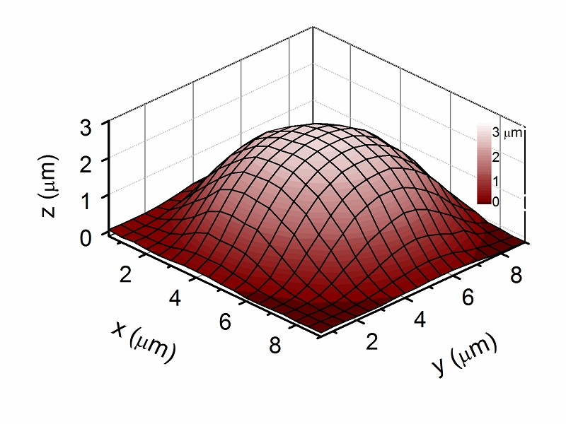
topographical map of an RBC adhered to a PLL-coated petri dish (map obtained in physiological solution at ambient pressure; lateral step: 500 nm).
Description
Numerous diseases, including atherosclerosis, hypertension, osteoporosis, muscular dystrophy, myopathies and cancer, are characterized by altered cellular response to mechanical forces. Cytomechanical studies have therefore the potential to contribute to understanding the mechanisms of these pathologies.
Cells elastic properties are investigated measuring the relationship between the force applied and the deformation induced. Beside whole-cell approaches, some techniques have been used to test local cell elasticity with a submicrometric spatial resolution. Among these, Scanning Ion Conductance Microscopy (SICM) allows one to investigate local elasticity in non-contact mode in living cells in vitro. A topographical scan can be carried out before measuring elasticity, to better control the area to be sampled. With SICM mechanical forces down to few tens of pN can be applied on a surface area smaller than 1 square micrometer, while measuring membrane indentations with resolution of tens of nm along z. Cell indentation was found to be reversible in a range of forces from few pN to about 1 nN, indicating that an elastic regime is investigated, with negligible visco-elastic effects.
We studied, among the others, the elastic properties of erythrocytes. These cells exhibit an easily reversible deformability that enables them to sustain the mechanical stress associated with the circulation, in particular through narrow capillary vessels. Differently from the behavior observed in other cells, we have found that the indentation of human erythrocytes does not grow linearly with the force applied and the apparent stiffness of the cell progressively increases for increasing force. Further studies are in progress to investigate possible effects on the treatment of microvascular dysfunctions.
Other scanning probe techniques available in our laboratory, for instance the atomic force microscopy (AFM), are useful for two main reasons. Firstly, working with spheres instead of standard sharp tips, one can measure in an alternative way the elastic modulus of the cells, verifying independently the data obtained via SICM. Additionally, the above mentioned techniques allow studying both the morphology and the elasticity of the substrates where the cells are grown.
INO Staff
Dinelli FrancoPellegrino MarioTognoni Elisabetta (Contact Person)