
EXPANSION-PALM of thick samples
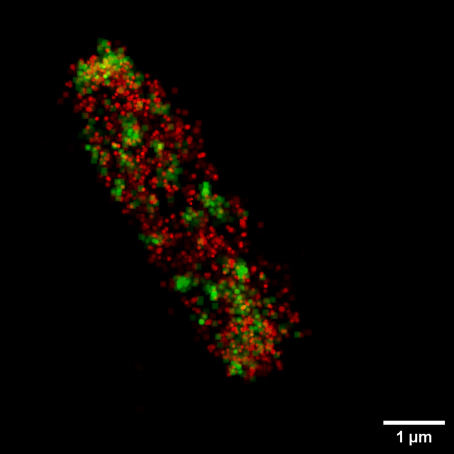
E.Coli Bacteria expanded 4 times with hisH-PamCherry and hisF-PAGFP. They are enzymes of the Histidine metabolic pathway we want to see whether they co-localize or not.
Description
In recent years we’ve been working on the combination of super-resolution PALM with Expansion microscopy. Expansion Microscopy allows to increase isotropically the dimension of the sample up to 4 times, thus in combination with high resolution imaging could in principle allow to reach the nanometer resolution. This technique is applied to the study of molecular mechanism involved in bacteria metabolism in collaboration with the group of Prof. Renato Fani at the Department of Genetics, as a proof of concept. Bacteria are of particular interest for this kind of application since they are very difficult to image with “traditional” super-resolution microscopy due to their very confined dimensions (1-2 microns in length, half a micron thick) which make it difficult to discriminate separate photoswitching chromophores, in particular when performing 3D super-resolution through astigmatism. Notably, excitation through highly inclined light sheet allows state of the art super-resolution in thick gels (Gardini et al. “Optimization of highly inclined illumination for diffraction-limited and super-resolution microscopy.” Optics Express 2023; Vignolini, et al. “Highly inclined light sheet allows volumetric super-resolution imaging of efflux pumps distribution in bacterial biofilms.” Scientific Reports 2024) and, in combination with Expansion Microscopy, allows measuring intermolecular distances down to 5 nm (article in preparation).
INO Staff
Gardini Lucia (Contact Person)
Personnel not belonging to INO
Chiara Caldini, Phd
Prof. Marco Capitanio
